 EN
EN
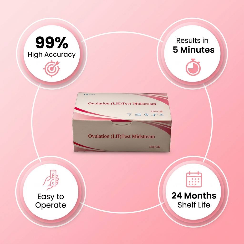
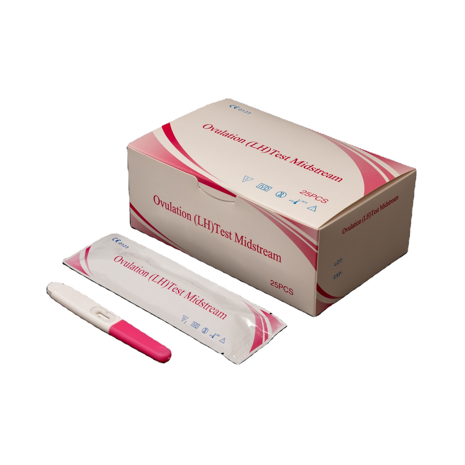
Ovulation Test Kit Working Principle and Hormone Detection Explained.
From an engineering standpoint, an ovulation test kit is one of the more elegant diagnostic tools in the POCT category. The device does one specific job: detect a transient hormonal surge in urine with enough sensitivity and speed to be clinically useful at the point of care. But when you look beyond the simple two-line readout, what you find is a precisely designed immunochromatographic system that bridges reproductive endocrinology with lateral flow assay technology. This article walks through the working principle step by step: what the kit is actually measuring, how the antibody-antigen interaction generates a visible signal, and what specification ranges matter most when evaluating an ovulation test kit for clinical or distribution use.
What Is LH and Why Does Its Detection Matter?
Luteinizing Hormone (LH) is a glycoprotein produced and released by the anterior pituitary gland. Under normal cycle conditions, LH levels remain relatively low throughout most of the menstrual cycle. Then, in response to rising estrogen levels from the developing follicle, the hypothalamus and pituitary trigger a sharp, brief surge — typically lasting 24 to 48 hours. It is this LH surge that directly signals the dominant follicle to complete egg maturation and release, with ovulation generally occurring within 24 to 36 hours of the surge peak.
In the context of fertility testing, this makes LH a highly specific and time-sensitive biological marker. Unlike basal body temperature or cervical mucus changes, which respond to ovulation after the fact, the LH surge precedes egg release — giving users or clinicians a predictive, actionable window. Most ovulation test kits are designed to detect this surge in urine, where LH levels mirror blood concentrations with a lag of approximately 3 to 6 hours.
That said, a common misconception needs addressing: many people believe ovulation occurs exactly 24 hours after a positive OPK result. In practice, research data suggests ovulation occurs anywhere within 24 to 36 hours of the detected surge, and individual variation in LH surge duration (some women experience a short surge, others a prolonged wave) means that a single threshold measurement does not tell the complete story. The test captures the beginning of the fertile window, not a precise countdown clock.
How the Ovulation Test Kit Detects LH — The Immunochromatographic Mechanism
The detection technology used in standard ovulation test strips is lateral flow immunochromatography — the same platform used in pregnancy tests, COVID-19 rapid antigen tests, and many other point-of-care diagnostics. Understanding this mechanism is essential for anyone evaluating, distributing, or sourcing these kits, because it directly governs what variables affect performance.
Step 1: Sample Application and Lateral Migration
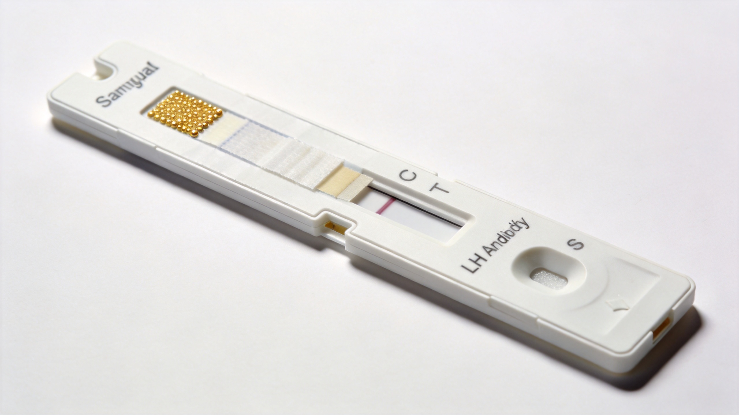
When a urine sample contacts the sample pad at one end of the test strip, capillary action draws the liquid laterally across a nitrocellulose membrane. This porous membrane forms the core of the detection system, with a precisely controlled pore structure that governs how fast the sample migrates and how efficiently analytes interact with the immobilized reagents along the way. The migration happens spontaneously, without any pumps or external equipment, which is what makes the lateral flow format ideal for home use and field diagnostics.
Step 2: Antibody-Antigen Binding in the Conjugate Zone
As the migrating sample reaches the conjugate pad, it rehydrates a pre-dried deposit of detection antibodies conjugated to colored nanoparticles (typically colloidal gold or colored latex microspheres). These labeled antibodies are specific to LH: they bind selectively to the LH molecule present in the urine sample, forming antibody-LH complexes that continue migrating with the sample flow toward the test zone.
Ovulation test kits use a sandwich immunoassay format for LH detection. LH is a relatively large glycoprotein (molecular weight approximately 30 kDa), which means it carries multiple epitopes on its surface, meaning distinct antibodies can simultaneously recognize different binding sites. This makes the sandwich format viable: one antibody grabs one part of the LH molecule, and a second antibody captures a different part, with the LH molecule in between.
Step 3: Signal Generation at the Test Line and Control Line
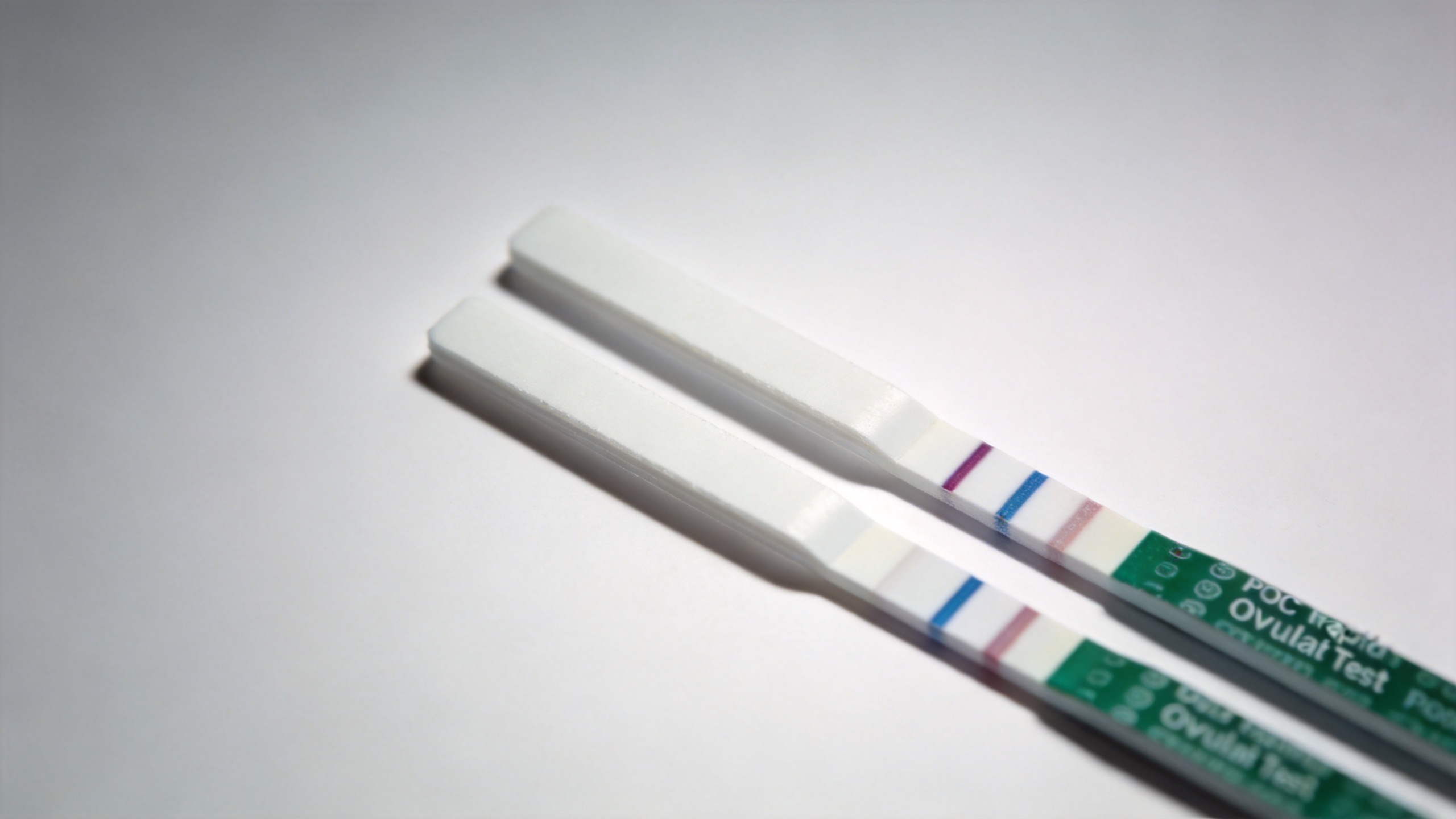
When the labeled antibody-LH complexes reach the test line zone, they encounter a second set of antibodies that are immobilized directly onto the nitrocellulose membrane at that location. These capture antibodies bind to the LH that is already attached to the labeled particle, completing the sandwich. The colored nanoparticles accumulate at the test line, producing a visible colored band. The darker and more distinct the test line, the higher the LH concentration in the sample relative to the kit's threshold.
A second line — the control line — positioned downstream. It contains anti-species antibodies that bind to any labeled antibody that has migrated through, regardless of whether LH was present. The control line serves as a procedural validity indicator: it confirms that sufficient sample volume was applied and that the reagents are functional. A valid test should show a control line. If no control line appears, the test result is invalid.
Understanding the Detection Threshold
One of the most practically important specifications in LH hormone detection is the threshold — the minimum urinary LH concentration that triggers a positive result. Most commercially available ovulation test kits use a threshold in the range of 20 to 50 mIU/mL, with many standardized around 25 mIU/mL. Research data indicates that beginning testing at cycle day 7 with a 25–30 mIU/mL threshold offers a positive predictive value of approximately 50–60% for ovulation within 24 hours, and a negative predictive value of around 98%.
If the threshold is set too high, the kit risks missing the surge in women who have a low-amplitude LH peak — a physiologically common variation that does not indicate an ovulation disorder. If it is set too low, the kit may generate false positives against the relatively elevated baseline LH seen in women with polycystic ovary syndrome (PCOS) or peri-menopausal hormonal shifts. This means the detection threshold is not just a manufacturing specification — it is a clinical positioning decision, and it matters when selecting which kit format to distribute or recommend for a specific user population.
Key Quality Parameters for Fertility Testing Procurement
When evaluating ovulation test kits from a diagnostic method and procurement standpoint, sensitivity (LH threshold) is only one dimension. A multi-parameter evaluation typically includes:
- Cross-reactivity profile: LH shares structural homology with FSH, hCG, and TSH. A well-characterized kit should document that its monoclonal antibody pair does not produce false-positive cross-reactions with these hormones at physiologically relevant concentrations.
- Analytical specificity: The degree to which the antibodies discriminate between intact LH and its fragments or related subunits affects result reliability across different sample matrices.
- Reading window: Most lateral flow results are valid within a defined reading window (typically 3 to 10 minutes). Results read outside this window may show background color changes that do not represent true positives.
- Storage conditions and shelf life: Nitrocellulose-based strips are sensitive to humidity and temperature. Kits intended for distribution to tropical or high-humidity markets require validated stability data under those conditions, not just standard 25°C/60% RH data.
- Regulatory documentation: For IVD diagnostic distribution across different regions, CE marking, FDA clearance, or local health authority registration documents confirm that the kit's claimed sensitivity and specificity have been independently evaluated.
In paper specifications, two kits with similar listed sensitivity thresholds may perform quite differently in field conditions. The actual membrane quality, conjugate particle uniformity, and monoclonal antibody affinity constants are variables that are not visible on a product sheet but directly affect real-world result consistency.
Summary
An ovulation test kit achieves hormone detection through a well-established lateral flow immunochromatographic process: the sample migrates across a nitrocellulose membrane, the LH analyte binds to labeled antibodies in the conjugate zone, and the resulting complexes are captured at the test line by immobilized antibodies to produce a visible signal. The working principle depends on the sandwich immunoassay format, the specificity of the monoclonal antibody pair, and the calibration of the detection threshold — typically in the 20–50 mIU/mL range. For procurement professionals, medical device distributors, and clinical buyers, understanding these technical parameters enables more confident and defensible sourcing decisions when comparing ovulation test kits across suppliers and intended markets.